


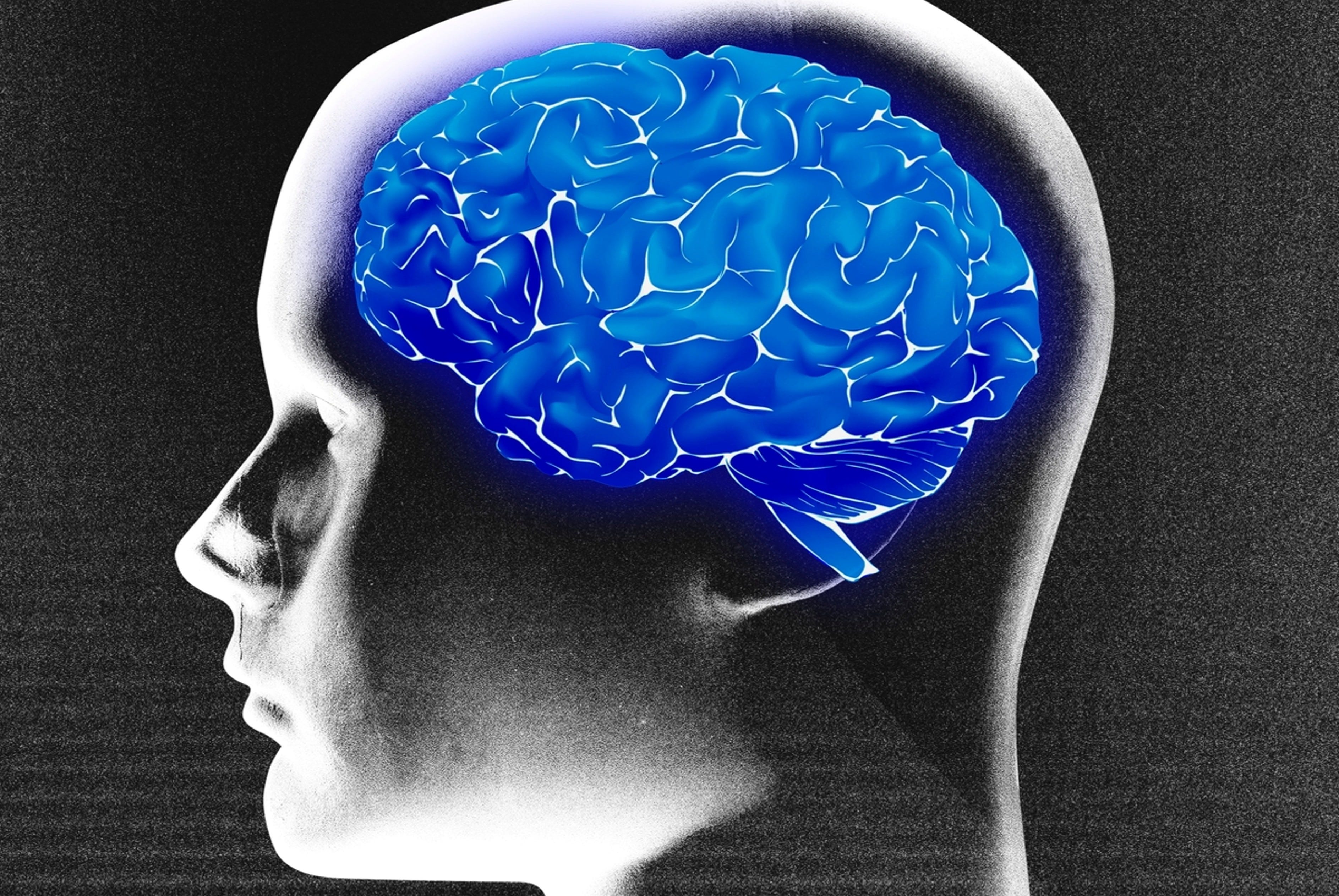
Flu Vaccine and Brain Health: What the Biological Evidence Says
It may do more than just prevent the neurodegenerative effects of influenza virus.

In my previous article, “Can Flu Shots Protect Your Brain Against Dementia? How Strong Is the Evidence?” I examined whether the flu vaccine could reduce the risk of dementia. I conducted an updated and targeted meta-analysis, synthesizing the most recent studies while adjusting for a key confounder — the healthy vaccinee bias. The results suggest repeated flu vaccinations (at least four doses) can help reduce dementia risk.
But epidemiological association (study of disease in populations) can only go so far. In this follow-up, I turn to the biology: what mechanisms might explain how the flu vaccine could protect the brain over the long term?
Methods
I used the same keywords to scan PubMed, the leading biomedical literature database, as I did in my previous article. The search returned 151 results as of April 10, 2025. After scouring them, I found four relevant studies examining the biological mechanisms underlying the neuroprotective effects (or lack thereof) of flu vaccines against dementia-related pathology.
These studies are as follows, sorted chronologically:
Li et al. (2019), China. Brain Research Bulletin.
Yang et al. (2020), China. Journal of Neuroinflammation.
Demuth et al. (2023), Germany. Frontiers in Pharmacology.
Cousins et al. (2023), UK. Journal of Neuroinflammation.
These studies are all published in reputable journals. All but one study involved animal models, which are the standard method to investigate biological causation since scientists can directly intervene with animal biology and observe the effect, although animal research ethics can be questionable. The same thing can’t be done in humans due to obvious ethical reasons. We can’t just cut open a living human’s brain, for instance. Because an epidemiological association exists, it also gives us a premise to look into the biological evidence, even if they were done with animal models.
After reading and synthesizing them narratively, I categorized the narrative into three themes and some caveats in this article.
Theme 1: Preventing Influenza’s Neurotoxic Effects
The most obvious neuroprotective mechanism of flu vaccines is arguably their ability to prevent the complications of influenza virus infection, which include neuroinflammation and neurodegeneration.
In a 2023 study, “Influenza vaccine is able to prevent neuroinflammation triggered by H7N7 IAV infection,” Demuth et al. infected mice with H7N7 influenza virus, which led to a cascade of damaging effects in the brain’s hippocampus: a surge in pro-inflammatory cytokines (TNFα, IL-6, and IFNγ), overactivated microglia, and loss of neuronal synaptic connections. Microglia are the brain’s immune cells that generate inflammation, which, while necessary to control infection, can damage neurons when excessive.
So far, this finding aligns with prior studies, where other influenza strains like H5N1 and H1N1 have also been found to trigger microglial activation and neuroinflammation in the brain, including the substantia nigra involved in Parkinson’s disease (PD) and hippocampus in Alzeimer’s disease (AD).
Demuth et al. then showed that when mice were vaccinated (with an inactivated vaccine) before infection, many of these harmful neurological effects were blunted or prevented altogether.
For instance, they used 3D imaging technology to reconstruct individual microglial cells in the mouse hippocampus. By stacking thin microscopic images and using specialized software, they created detailed models showing cell size, branching complexity, and how many neuronal fragments were engulfed by the microglia. This allowed the researchers to see how flu infection and vaccination affect microglial behavior.
With this technology, Demuth et al. showed that influenza infection increased the microglial cell volume, soma size, and branch points in unvaccinated mice compared to vaccinated mice (Figure 1). Each of these changes is a hallmark of microglial activation. When microglia detect threats like viral infection, they undergo physical changes: their soma (cell body) swells, cell volume increases to accommodate increased activity, and their branches expand as a sign of heightened alertness. But vaccination dampened these changes, keeping microglia closer to their resting state.

Next, Demuth et al. reconstructed 3D images of microglial digestive compartments called lysosomes (labeled with LAMP-1) and neuronal synaptic terminals (labeled with Homer-1). This allowed the researchers to visualize how microglia interact with neurons during influenza-induced brain inflammation. Specifically, they tracked whether microglia were engulfing the synaptic terminals of neurons by seeing if Homer-1-positive synaptic material was present inside LAMP-1-positive lysosomes. This is crucial because microglial ‘eating’ of synapses is a well-known mechanism of synaptic loss in neurodegenerative diseases.
By visualizing this process, the study provided evidence that influenza infection increased the lysosomal engulfment of synaptic components by microglia, suggesting that influenza triggers microglia to strip away neuronal connections. But flu vaccination prevented this synaptic engulfment to some extent, helping to preserve the neuronal health (Figure 2).
So, these findings suggest that the flu vaccine may protect against influenza-induced neuroinflammation and synaptic loss. By keeping microglia in a less activated state, vaccination prevents excessive pruning of neuronal connections that would lead to overt neurodegeneration.

Theme 2: Modulating the Brain’s Immune Response
Keep reading with a 7-day free trial
Subscribe to The Infected Neuron to keep reading this post and get 7 days of free access to the full post archives.