


What If Something Else Lives in Your Brain? The Science and Secrets of Brain Microbiome
What it is and why it matters.

This article was originally published at Microbial Instincts on May 14, 2024.

What if there are unseen inhabitants within your brain?
In 2020, I wrote an article titled “Is There A Brain Microbiome Now?” to delve into this question. I described preliminary findings in 2018 when scientists captured images of microscopic rod-like structures — likely bacterial — in human brain samples preserved shortly after death. This shocked the scientific community, as the brain is believed to be a sterile organ.
Four years later, this topic crossed my mind again. I wondered why I hadn’t heard about it from the popular media or academia. Curious, I searched PubMed — the leading biomedical literature database — about the brain microbiome. The search returned 36 papers as of 14 May 2024, which is very few compared to, say, the gut microbiome, with over 20,000 papers. No wonder I hadn’t heard about the brain microbiome.
Of the 36 papers, 26 are about the gut-brain axis. This axis refers to the influence of the gut microbiome on the brain, which does not involve any brain microbiome. This leaves only 10 or fewer relevant papers, and reading them has been very interesting. So, let me tell you about the science and secrets of the fascinating brain microbiome.
Author’s note: This article may be complex to read, so please feel free to jump to the last two sections for the main takeaways.
Part I: The first hint
The possible existence of a brain microbiome was first hinted at by a 2013 study from Canada, where scientists were investigating whether brain damage seen in HIV/AIDS patients involves microbial invasion.
Using advanced genetic techniques, they found genetic material from 173 different species of bacteria and viruses in the brain samples, specifically in glial cells of the brain white matter.
The most dominant bacterial group was proteobacteria (Figure 1), while viruses include bacteriophages and herpesviruses. What’s interesting is that both HIV-infected and uninfected brain samples showed similar microbial patterns (Figure 1), suggesting that microbes could be living in our brains even if we’re not immunocompromised by HIV/AIDS.

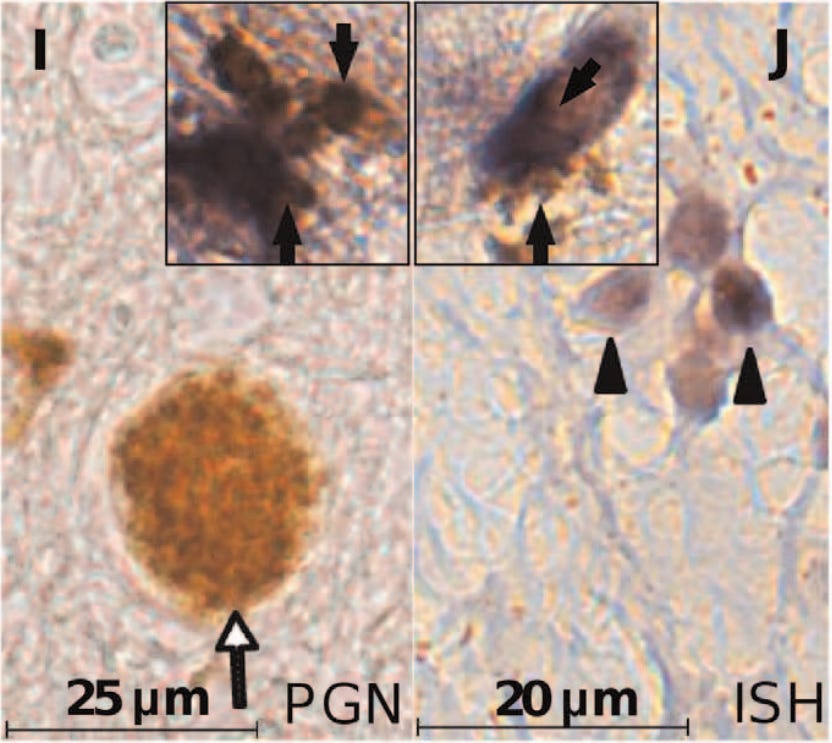
The findings were confirmed using various methods that detect bacterial genes and structures, including staining for peptidoglycan (a component of bacterial cell wall) in the brain samples (Figure 2). The staining patterns were also consistent with how a bacteria would look like.
The study went further by transplanting human brain tissues into immunocompromised mice. After 7 weeks, a similar bacterial profile dominated by α-proteobacteria was observed in the mice's brains.
In a final ingenious move, they repeated the experiment with brain tissues that were heat-treated to kill bacteria, and they found no bacterial presence in the mice’s brains afterward— suggesting that the bacteria found in the human brain samples were alive and active.
“Thus, α-proteobacteria represented the major bacterial component of the primate brain’s microbiome regardless of underlying immune status,” the study authors concluded, “which could be transferred into naïve hosts leading to microbial persistence in the brain.”
“While this early study used relatively small sample sizes, it provided the types of validation studies needed to counter the common assumption that the brain is sterile,” US-based neuroscientist Dr. Christopher D. Link wrote in a 2021 review paper in response to the 2013 study.
If distinct microbial profiles are identified in different brain regions, it suggests that these microbes are not random contaminants but are specific to certain areas.
Part II: Possible contamination
Dr. Link’s 2021 review paper, “Is There a Brain Microbiome?” also questions the existence of the brain microbiome by raising the possibility of contamination from sources such as laboratory reagents, extraction processes, and residual blood in tissue samples.
However, the 2013 Canadian study did consider and address the possibility of sample contamination in their experiments. They consistently used controls for all the chemicals and reagents at every step of their genetic analyses, even repeating the entire process in a different lab and staff. The brain tissues examined came from various sources — some were obtained from autopsies 12 to 24 hours after death, while others were taken from surgery in a sterile environment and immediately frozen.
A valid experiment must have the appropriate control groups to ensure that the results observed are not due to external factors, such as reagent contamination). For example, reagent controls may involve running the experiment with the reagents alone; if no bacterial genes were detected, we can eliminate the possibility of reagent contamination.
But a 2021 study from Germany might have put the brain microbiome science to rest. The study’s title paints a gist of what it’s about: “Much ado about nothing? Off-target amplification can lead to false-positive bacterial brain microbiome detection in healthy and Parkinson’s disease individuals.”
This study used similar genetic techniques to find bacterial sequences in brain samples from healthy people, Parkinson’s disease patients, and mice. While they did find bacterial genes in all brain samples, the amount of bacteria was meager. Further analyses designed to analyze contaminants showed that these bacterial signals were likely due to DNA contamination or off-target sequencing of host DNA that’s non-bacterial.
In gene sequencing, primers are designed to bind to specific gene sequences to flag them for sequencing. But sometimes, the primers may bind to off-target areas (e.g., a part of the human genome) with similar gene sequences to the target of interest (e.g., bacteria). Such off-target is more likely to occur when the amount of untargeted genes (human genes) far outweighs the targeted genes (bacterial genes) in the sample.
As the authors of the German study wrote, “Off-target amplicons probably occur due to the extremely low bacterial biomass in samples and primer competition with the dominant host DNA background.”
The brain microbiome, especially at the frontal lobe, may evolve from a healthy state (proteobacteria-dominant) to a disease state of AD (actinobacteria-dominant).
Part III: Maybe it’s not just contamination
The 2021 German study may have dampened the enthusiasm for further research in brain microbiome science. It wasn’t until late 2023 that an impactful study from the US revived the topic.
One of the study’s senior authors is Jeffry R. Lapides, PhD., an adjunct professor at Drexel University College of Medicine. I felt a mix of surprise and familiarity upon seeing a new study from Dr. Lapides. I exchanged a few emails with him about the brain microbiome years ago, as well as recently when I reached out to him about his new research.
This latest study initially sought to characterize the pathogenic brain microbiome of patients with Alzheimer’s disease (AD). While the brain is believed to be sterile, the blood-brain barrier can weaken with age or neurodegenerative diseases, allowing pathogens to leak into the brain.
As a result, various pathogenic viruses and bacteria have been found in the brains of patients with neurodegenerative diseases like Alzheimer’s and Parkinson’s. (I wrote about this in detail before here and here.)
While the 2023 study did find a pathogenic microbiome profile in the brains of AD patients, it also found a distinct microbiome in the brains of control patients without AD. Specifically:
AD brains have more abundant Cutibacterium acnes, Staphylococcus epidermidis, Acidovorax ebreus, Acinetobacter tjernbergiae, Acidovorax temperans, Noviherbaspirillum soli, and Methylobacterium goesingense, with the most notable change being C. acnes (an actinobacterium).
Control brains have more abundant Acinetobacter junii, Comamonas jiangduensis, Cloacibacterium normanense, Pseudomonas putida, Pseudomonas thermotolerans, and Diaphorobacter nitroreducens, with the most notable change being A. junii (a proteobacterium).
(Note: While the genus of A. junii is Acinetobacter, it doesn't belong to the actinobacteria phylum, even though they may sound similar).
Such distinct brain microbiome profiles are similar to a prior British study in 2017, which also found the presence of more abundant actinobacteria in the brains of AD patients, although the abundance of proteobacteria was similar in both AD patients and control individuals.
Based on these results, further computational analyses mapped five distinct brain microbiome profiles, each correlated with varying degrees of AD prevalence (Figure 3). For example, a brain microbiome profile marked as 0% AD (green) indicates that these samples belong to individuals with no signs of AD. Conversely, a profile labeled as 88% AD (magenta) indicates that 88% of the samples belong to those with AD.

It gets more interesting.
The study took a step further by analyzing its results based on the different brain regions—i.e., frontal lobe, temporal lobe, and entorhinal cortex—from which the samples were collected. These brain regions are involved in cognitive processes and often degenerate in AD.
Apparently, the magenta samples — indicative of the brain microbiome profile of AD — are more likely derived from the frontal lobe rather than the temporal lobe or entorhinal cortex among AD patients (Figure 4).
“Looking at this, it was obvious what was going on. Magenta samples appear at least once in almost all the AD samples and almost never in the controls,” Dr. Lapides told me in an email. “Magenta [significantly] predicts AD, which, of course, does not explain why.”
In other words, while the study found that the magenta-colored brain microbiome profile reflects AD, the mechanisms behind this association remain unknown. We'll discuss this further in the next section.
That said, this small set of data, the authors wrote, suggests “that subjects with the magenta class in their frontal lobe are more likely to have the diminished cognitive abilities that result in AD diagnoses.”

But is it contamination again? No, this study also performed careful analyses to rule out such a possibility. For instance, they excluded samples with low bacterial yields to minimize the occurrence of off-target sequencing. They also included reagent controls without any biological samples in their experiments, as well as performed similar computational analyses of contaminants as the 2021 German study.
What’s more convincing, though, is that the study identified different microbiome profiles in different parts of the brain.
If distinct microbial profiles are identified in different brain regions, it suggests that these microbes are not random contaminants but are specific to certain areas. This specificity could imply a functional or ecological significance, which is less likely to be due to contamination.
In other words, blood contamination or errors in DNA amplification would affect all samples in a similar manner, leading to a more uniform microbial distribution across the brain. The presence of different microbial communities in different brain areas reduces the likelihood that these findings are solely due to contamination or off-target sequencing.
The presence of different brain microbiome profiles in distinct brain regions was first demonstrated in a 2020 study from the US, which found different microbial diversity between the hippocampus and cerebellum in the brains of AD patients. But this study did not perform the necessary analyses to rule out contamination or off-target sequencing, so I didn’t discuss it in depth. That said, at least this study supports the existence of varying microbiome profiles in different brain areas.
“We described spatiotemporal changes in these microbiomes from microscopic to macroscopic spatial scales,” authors of the 2023 study concluded. “Specifically, we uncovered an evolving microbiome in the human brain that begins, perhaps as a healthy microbiome, and then gradually changes until it is unquestionably associated with [AD].”
The idea that a microbiome might play a role in brain conditions like AD suggests we could one day treat these diseases with microbial interventions, much like how we use probiotics for gut health.
Part IV: Recap and some caveats
The field of brain microbiome is still in its infancy, with only a handful of studies supporting its existence. The strongest evidence comes from the 2023 American study, which showed that the brain microbiome, especially at the frontal lobe, may evolve from a healthy state (proteobacteria-dominant) to a disease state of AD (actinobacteria-dominant).
This 2023 study is further supported by at least 3 previous studies:
2013 Canadian study: Proteobacteria dominate the brain microbiome of individuals regardless of immune status.
2017 British study: Actinobacteria are more abundant in the brains of AD patients, with about the same abundance of proteobacteria in both AD patients and control individuals.
2020 American study: Brain microbiome profiles differ between the hippocampus and cerebellum regions in the brains of AD patients.
While we already ruled out the possibility of contamination and off-target sequencing in part III, a few major caveats must be discussed.
First, only the 2013 Canadian study examined the brain microbiome of young individuals. Because AD primarily affects older adults, the control groups in the other studies are also of advanced age. However, the blood-brain barrier weakens with age, which may allow microbes to infiltrate the brain more easily. Thus, it’s difficult to rule out the potential confounding effects of old age in influencing the study results.
Second, these studies only uncovered pieces of microbial genetic evidence and images of bacteria structures from the brain samples. But definitive evidence would come from culturing these microbes alive. However, it’d be unfair not to acknowledge the challenge of culturing microbes outside their natural habitat. Microbes residing in a microbiome may rely on specific organ physiology and other microbes to survive — conditions that may not be replicable in the lab. In fact, up to 70% of the gut microbiome has never been cultured alive in the lab.
Third, we don’t really know what roles the brain microbiome plays. These studies only identified the existence of a brain microbiome. But we can infer certain roles based on extrapolation of existing knowledge.
For instance, actinobacteria, particularly species like C. acnes—frequently identified as part of the brain microbiome of AD patients—are known to elicit inflammatory responses. In AD, these bacteria could exacerbate inflammatory processes in the brain, which drives oxidative stress and the formation of neurotoxic amyloid plaques.
In contrast, proteobacteria like A. junii —frequently identified in the normal brain microbiome— are found in the environment, where they break down organic materials and pollutants, contributing to nutrient cycling processes. But A. junii can also cause urinary tract infections in humans in rare cases. Aside from that, the existing literature doesn’t have any data on what A. junii has to do with brain physiology.
Now, would the existence of a brain microbiome redefine our understanding of neurobiology?
Part V: Why it matters and the next steps
The idea that a microbiome might play a role in brain conditions like AD suggests we could one day treat these diseases with microbial interventions, much like how we use probiotics for gut health.
A recent study from the US, China, and Japan used a probiotic’s bacteriocin — called nisin — to prevent adverse changes in the brain microbiome and the development of AD in mice. Specifically, this study used oral infection to stimulate periodontal diseases in mice, which altered their brain microbiome and triggered AD. Periodontal diseases are a known risk factor for AD because oral health is closely tied to brain health.
However, oral treatment with nisin prevented this alteration by preserving the abundance of the proteobacteria phylum and Acinetobacter genus in the brain microbiome of mice. As discussed above, such microbial patterns are consistent with what a healthy brain microbiome would look like in humans. Such consistency is also a testament to the reliability and validity of the data from these studies.
Based on the findings, the authors “hypothesized that nisin could prevent the periodontal pathogen-mediated neurodegeneration as a direct consequence of its ability to reverse the changes in the brain microbiome, immune profiles, and pathologic protein deposition.”
The existence of brain microbiome is also consistent with recent trends in neurobiology, where pathogenic viruses are increasingly recognized as significant drivers of neurodegenerative diseases. I wrote about this in detail before in “The Viral Origin of Alzheimer’s Disease Remains Undecoded. But What We’ve Seen So Far Is Worrying” and “The Case for Virus Origin of Neurodegenerative Diseases Is Getting Stronger and More Important.”
To move forward, we need a better understanding of the brain microbiome, which is still a black box full of secrets.
Key questions include:
Does the brain microbiome differ based on age and sex?
Does it come from the gut microbiome or somewhere else?
What mechanisms allow these microbes to survive and function within the brain’s supposedly sterile environment?
Does it interact with the brain, or is it just an inactive entity?
Does it play a role in the development of neurological diseases?
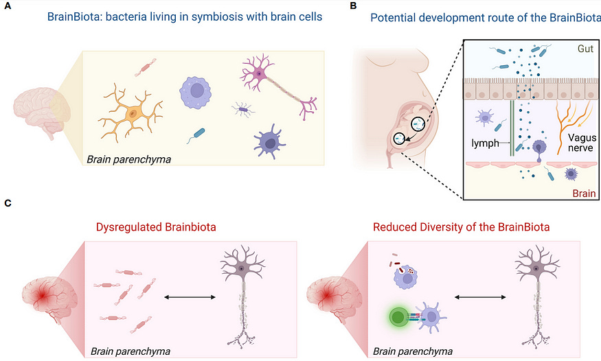
In a 2022 review paper, scientists from Denmark and Germany theorized that the brain microbiome may originate from the gut microbiome during early fetal development. They also suggest that the brain microbiome may play important immunomodulatory roles in helping to prevent autoimmunity and chronic inflammation (Figure 5).
The last two points are particularly relevant in clinical medicine. For instance, if certain bacteria are found to influence the immune response or interact with neural pathways in the brain, these could become targets for therapeutic intervention of neurological diseases.
Personally, I am both excited and cautious about the potential of this research. The brain is our most complex organ, and the thought of adding a microbiome layer to our understanding of it is fascinating.
The discovery of the gut microbiome revolutionized the field of gastroenterology and how we view gut health. Similarly, insights into the skin and oral microbiomes are reshaping our approaches to dermatology and dental health. Now, would the existence of a brain microbiome redefine our understanding of neurobiology?
If you have made it this far, thank you. If you enjoyed this, please subscribe below and share it with others. You can also tip me here. :))
Very excited to read this! I studied microbiology, and the microbiome was undoubtedly the most fascinating area to learn about - especially how it is involves in such diverse diseases and treatments =)